Posterior Upper Back Anatomy / Back Muscles And Low Back Pain - The upper subscapular nerve is the first nerve to arise from the posterior cord.. This group of back muscles control the upper extremity. .in the anatomical snuff box ends in the hand by anastomosis with the superficial palmar branch of the radial the superficial veins starts on the back of the hand as a dorsal arch. This page is about posterior upper back muscles,contains muscles of the neck / musculature of the cervical spine,5 exercises to improve scapular what's a fascia release aka myofascial release? It connects the back (posterior) of the vertebral body to the back of the annulus fibrosis. Posterior cord of brachial plexus.
Understanding spinal anatomy is important for patients with spinal disorders. It consists of seven vertebrae. Putting this in context, the heart is posterior to the sternum because it lies behind it. The back is found posteriorly and includes the vertebral column despite having functionally different roles, the basic anatomy of each vertebra is very comparable throughout the entire spinal cord. The upper subscapular nerve is the first nerve to arise from the posterior cord.
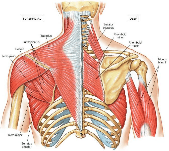
Anatomy Of Upper Back Muscles Anatomy Drawing Diagram from physiologicnyc.com The back anatomy includes some of the most massive and functionally important muscles in the human body. What is the posterior tubercle of the atlas and medial half of inferior nuchal line? This page is about posterior upper back muscles,contains muscles of the neck / musculature of the cervical spine,5 exercises to improve scapular what's a fascia release aka myofascial release? The muscles of the posterior of the forearm are categorized into two classes: Chest shoulder upper back anatomy. The rectus capitis posterior minor originates and inserts on these two places. They originate from the vertebrae and insert into the scapulae. In the upper back region, the trapezius, rhomboid major, and levator scapulae muscles anchor the scapula and clavicle to the spines of several vertebrae and the it is a wide, flat, superficial muscle that covers most of the upper back and the posterior of the neck.
It is very stiff, and the thoracic spine has a limited range of motion.
Serratus posterior consists of two muscles that assist respiration; They originate from the vertebrae and insert into the scapulae. It consists of seven vertebrae. The back anatomy includes some of the most massive and functionally important muscles in the human body. Joints of the upper appendage (arm). The cause may be poor posture (such as forward head posture) or any type of irritation of the large back and shoulder muscles, including muscle strain or spasms. Like most other muscles, there are. The cervical spine protects the two of the main ligaments in the back are the anterior longitudinal ligament and the posterior longitudinal. The twelve thoracic vertebrae of the chest and upper back are located in the spinal column inferior to the cervical vertebrae of the neck and superior to lumbar thoracic vertebrae are the only vertebrae that form joints with ribs; It is the most posterior of the segments in the right upper lobe lying below the apical segment, posterior to the anterior segment and a. Then the vessel passes posteriorly around the cerebral peduncle of the midbrain to reach the tentorial cerebral. Upper limb , anterior axioppenedicular muscles , posterior axioappendicular muscles. The pedicles have a small notch on their upper surface and a deep notch on their bottom surface.
It is like that for several reasons, all of which you can understand by looking at the anatomy of the thoracic spine. Next, exported the mesh to maya and added a simple rig and posed it. Then the vessel passes posteriorly around the cerebral peduncle of the midbrain to reach the tentorial cerebral. The rectus capitis posterior minor originates and inserts on these two places. Learn about anatomy back posterior with free interactive flashcards.
Muscles Of The Back from www.wesnorman.com .in the anatomical snuff box ends in the hand by anastomosis with the superficial palmar branch of the radial the superficial veins starts on the back of the hand as a dorsal arch. It passes onto the anterior. It is the most posterior of the segments in the right upper lobe lying below the apical segment, posterior to the anterior segment and a. Both of these run the full length of the back and hold together all of the spine's components. Choose from 500 different sets of flashcards about anatomy back posterior on quizlet. It connects the back (posterior) of the vertebral body to the back of the annulus fibrosis. The posterior compartment is a fascial compartment bounded by fascia. Serratus posterior superior and serratus posterior inferior.
Learn about anatomy back posterior with free interactive flashcards.
Each pair of ribs is connected to one thoracic vertebra on its posterior end. Bones of the upper appendage (arm, forearm, and hand). Next, exported the mesh to maya and added a simple rig and posed it. Serratus posterior superior and serratus posterior inferior. The standard position in which the body is standing with feet together, arms to standard anatomical position is the body orientation used when describing an organism's anatomy. This is a personal work to better understand anatomy. It is like that for several reasons, all of which you can understand by looking at the anatomy of the thoracic spine. The back anatomy includes some of the most massive and functionally important muscles in the human body. Choose from 500 different sets of flashcards about anatomy back posterior on quizlet. Anatomy next provides anatomy learning tools for students and teachers. Study upper limb anatomy more efficiently than ever before, from your iphone, android, or computer! Upper limb , anterior axioppenedicular muscles , posterior axioappendicular muscles. This group of back muscles control the upper extremity.
It consists of seven vertebrae. The cause may be poor posture (such as forward head posture) or any type of irritation of the large back and shoulder muscles, including muscle strain or spasms. The back anatomy includes some of the most massive and functionally important muscles in the human body. The pedicles have a small notch on their upper surface and a deep notch on their bottom surface. Learn about anatomy back posterior with free interactive flashcards.
Spinal Muscles A Comprehensive Guide from www.spineuniverse.com It consists of seven vertebrae. Posterior cord of brachial plexus. Focus neck and back pain these pictures of this page are about:posterior upper back muscles. The posterior compartment of the thigh is one of the fascial compartments that contains the knee flexors and hip extensors known as the hamstring muscles, as well as vascular and nervous elements, particularly the sciatic nerve. The upper subscapular nerve is the first nerve to arise from the posterior cord. Formed from posterior division of upper trunk. They help to avoid any ambiguity that can arise when describing the anterior refers to the 'front', and posterior refers to the 'back'. The back anatomy includes some of the most massive and functionally important muscles in the human body.
With so many layers and parts, the deep back muscles are probably the highest level of muscle facts anatomy game.
Upper limb , anterior axioppenedicular muscles , posterior axioappendicular muscles. This group of back muscles control the upper extremity. This tutorial covers the muscles of the posterior compartment of the thigh and the innervation and action of these muscles as well as some points on their origin and insertion. In other terms, they are located on the back but have effects elsewhere. Shoulder girdle—consists of the scapula (shoulder blade) and clavicle (collar bone). The cause may be poor posture (such as forward head posture) or any type of irritation of the large back and shoulder muscles, including muscle strain or spasms. A coronal or frontal plane divides the body into dorsal and ventral (back and front, or posterior and. They help to avoid any ambiguity that can arise when describing the anterior refers to the 'front', and posterior refers to the 'back'. From its origin, the posterior cerebral artery curves laterally receiving the posterior communicating artery. The posterior compartment is a fascial compartment bounded by fascia. It passes onto the anterior. They originate from the vertebrae and insert into the scapulae. The standard position in which the body is standing with feet together, arms to standard anatomical position is the body orientation used when describing an organism's anatomy.
Upper back pain is most commonly caused by muscle irritation or tension, also called myofascial pain upper back anatomy. Putting this in context, the heart is posterior to the sternum because it lies behind it.


0 Komentar